The in situ spatial transcriptomics workflow begins with sectioning tissue samples onto a Xenium slide, followed by sample preparation for probe hybridization, ligation, and amplification. The DNA probes used in this process also include gene-specific barcode sequences for transcript identification. When multimodal segmentation is applied, the tissue is additionally stained with markers targeting the cell interior, nucleus, and membrane. Notably, multiplexed protein detection is also possible within this workflow.
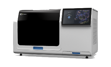
Within the Xenium Analyzer, multiple cycles of fluorescent probe hybridization, imaging, and signal decoding are performed. Final data visualization is carried out both on-instrument using the Xenium Analyzer and off-instrument using the Xenium Explorer software.
The platform enables single-molecule localization with high spatial precision—less than 30 nm in the lateral (XY) axis and approximately 100 nm in the axial (Z) axis.